

Coal mining has long been the backbone of industry, powering progress and innovation in our society. Amid this vital work, the threat of black lung disease casts a dark shadow over the lives and health of coal miners.
According to the American Lung Association, an estimated 16 percent of coal workers are affected by black lung.
Black lung is associated with prolonged exposure to respirable coal mine dust, inhalable dust consisting of airborne particles so tiny they can penetrate deep into the lungs and cause serious health problems. The disease often is identified through a miner’s work history and X-ray findings.
When the dust contains a lot of crystalline silica, it is especially hazardous. Workers with black lung disease often experience difficulty breathing, persistent coughing, and chest tightness caused by inflammation and scarring in their lungs. In recent years, the coal mining industry has implemented many safety measures in an effort to mitigate exposures.
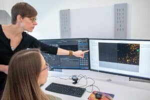
Emily Sarver, the Stonie Barker Professor of Mining and Minerals Engineering, has dedicated more than a decade to investigating respirable dust in the mine atmosphere using state-of-the-art electron microscopy tools. Now, she and her team are transferring their expertise to detect and analyze silica and other mineral particles in lung tissue with a goal to provide more insights on the outcomes of specific dust exposures, such as silicosis, a disease caused by inhaling silica dust.
“Black lung disease among coal miners remains a persistent issue, but there are a number of other industries where dust exposure monitoring is less pervasive, and we are starting to see outbreaks of silicosis among workers,” said Sarver. “By studying both occupational exposures and the dust burden in lung tissue, we can better understand disease development and the lung’s response over time.”
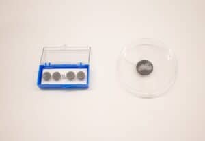
Using the electron microscope, Sarver and her team can see incredibly small details of lung tissue and study the types and sizes of dust particles in affected areas. With advanced hardware and software, they can automate the analysis to be much more efficient and limit any bias in interpretation of results. They recently demonstrated this approach by analyzing dust particles in lung tissue specimens from 10 coal miners with severe black lung disease. Moving forward, they aim to apply the research to workers in other dust-exposed occupations, such as construction and engineered stone fabrication.
Ultimately, Sarver wants to correlate what she sees in environmental samples with lung tissue analysis to better understand the histology and pathology of dust-related diseases. Although the traditional light microscope has long been used by pathologists, the electron microscope can magnify objects to resolve micron and even submicron particles, which are those small enough to make their way into the air-exchange region of the lungs. Additionally, the electron microscope can be used with X-ray spectroscopy to enable elemental analysis of particles and determine their mineralogy.
“We have had the opportunity to look at a limited number of tissue specimens, but the reality is that means we are looking at someone who has already been overexposed, and it is impossible to know everything about their history,” said Sarver. “But we could apply the same methods to study cause and effect in experiments that merely simulate human exposures.”
To better prevent occupational dust exposures, Sarver’s team also is working on an idea to use portable optical microscopy with automated image analysis to provide miners and employers with crucial information about the types and concentrations of airborne particles.
The concept would provide near real-time data that could enable quick and informed decisions to mitigate workers’ exposure to respiratory hazards, ultimately fostering a safer and healthier working environment. Real-time monitoring tools have long been sought for mine dust monitoring, and new regulations are only adding to the push for new solutions.
“We’ve done a lot with the electron microscope in the lab, but for a field application a simple light microscope, even a cell-phone microscope, is really appealing,” said Sarver. “We are envisioning a process whereby an airborne dust sample can be quickly collected, prepared, and imaged. An algorithm then applies a particle classification model to sort out the main types of particles present. This approach could allow proactive decision making to lower harmful dust exposures, and ultimately lower the number of occupational lung diseases.”
Sarver’s research on respirable dust has been funded by the Centers for Disease Control and Prevention and National Institute for Occupational Safety and Health through its Mining Research Division and the Alpha Foundation for the Improvement in Mine Safety and Health, and it’s been made possible through sampling agreements with numerous industry members and collaborations with other researchers at:
- University of Illinois Chicago
- National Jewish Health
- U.S. Geological Survey
- University of Calgary
- University of Colorado
The collaboration between mining engineers and medical researchers highlights the importance of interdisciplinary approaches in tackling complex health issues. This can lead to improved occupational health standards and practices globally.
By Hailey Wade